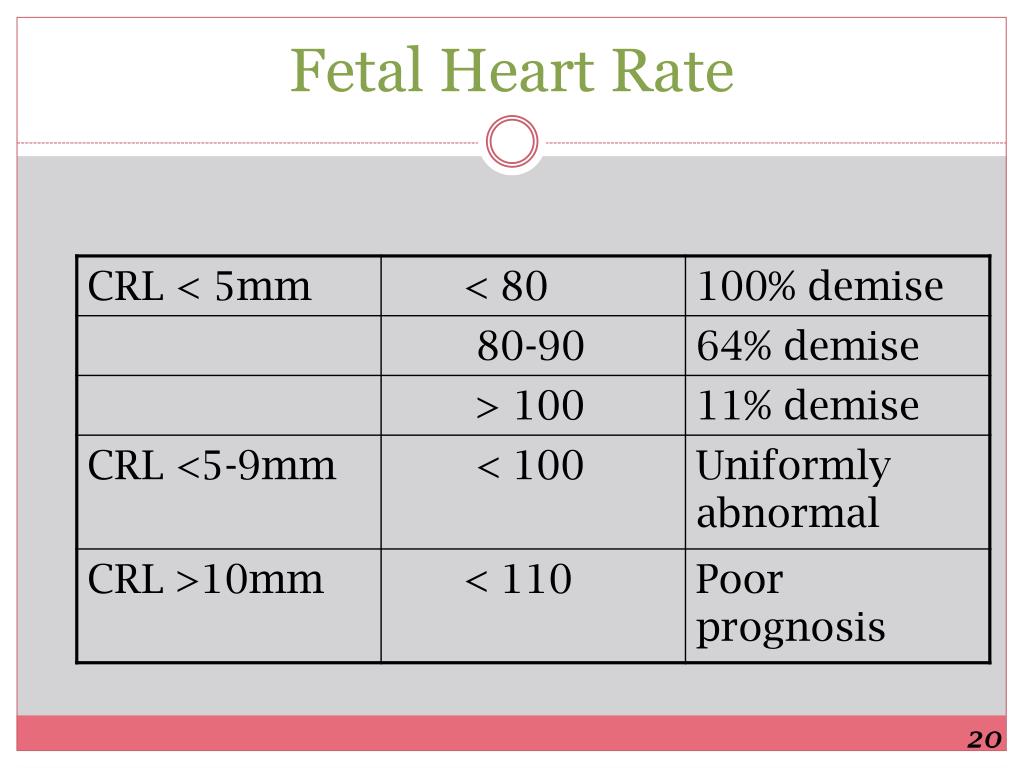


1 With this technique, which demonstrates the relationship between atrial contraction (blood flow reversal in the superior vena cava) and ventricular contraction (forward pulsatile flow in the ascending aorta), A–V and ventriculo‐atrial time intervals can be defined. More recently, Fouron and colleagues described the use of a simultaneous superior vena cava and ascending aorta Doppler assessment obtained in a sagittal view through the fetal thorax. Pulsed Doppler interrogation through the left ventricular inflow and outflow tract, with evaluation of the relationship between the a wave during atrial contraction and the outflow signal, can provide additional information although it is limited when atrial contraction occurs against a closed AV valve such as may occur in AV block. The quality of the assessment is significantly affected by image resolution and fetal position. In M mode assessment, a simultaneous recording of atrial and ventricular wall motion is acquired. The echocardiographic diagnosis of fetal arrhythmias involves the assessment of the chronological relationships between atrial and ventricular contractions, with assumptions about their electrophysiological relationships. Most centres currently rely on echocardiographic modalities to define the fetal rhythm which include M mode, and pulse and tissue Doppler (fig 1 1). While fetal electrocardiography has been described, the low p wave voltage, frequent inability to document heart rates above and below a normal range, particularly at rates that are similar to the mothers, limitations in fetal signal acquisition between 28–34 weeks, and use of signal averaging make assessment of the majority of fetal arrhythmias by electrocardiography at present not feasible or practical. This article will review the diagnostic modalities currently employed in the assessment of fetal rhythm disturbances, the types of arrhythmias encountered before birth, as well as the management and outcome of fetal arrhythmias with and without intervention.Īccurate diagnosis of rhythm and conduction abnormalities can be challenging given the lack of conventional ECGs. For such abnormalities prenatal diagnosis with the potential for prenatal, perinatal and neonatal intervention may be critical and may ultimately improve outcome. Less than 10% of referrals for fetal rhythm abnormalities have a sustained tachyarrhythmia or bradyarrhythmia considered to be of clinical significance, as they may indicate severe systemic disease or may have the potential to compromise the fetal circulation themselves. The vast majority of affected pregnancies have isolated premature atrial contractions which may have even spontaneously resolved by fetal echocardiographic assessment. While such clinical assessments begin at 12–14 weeks, most fetal arrhythmias are detected only after 20 weeks. They are usually identified by the obstetrical clinician who detects an abnormal fetal heart rate or rhythm using a Doppler “listening device” at routine assessment of the pregnant mother. In the healthy fetus the heart rate is regular, usually remains between 110 and 180 bpm, and has a beat‐to‐beat variation of 5–15 bpm.įetal rhythm abnormalities, which include fetal heart rates that are irregular, too fast or too slow, occur in up to 2% of pregnancies and account for 10–20% of the referrals to fetal cardiologists. By 20 weeks the average (SD) fetal heart rate is 140 (20) bpm with a gradual decrease to 130 (20) bpm by term. The rise in heart rate is followed by a decrease to 150 bpm by 14 weeks, likely as a consequence of increasing parasympathetic control and improved myocardial contractility. With further growth and maturation of the conduction system, including definition of the sinoatrial node as the primary cardiac pacemaker with its highest intrinsic rate of spontaneous depolarisation, there is a subsequent increase in the rate to 170 bpm by 9–10 weeks. By 5–6 weeks the normal mean fetal heart rate is 110 beats/min (bpm). While the exact timing of onset of the atrioventricular (AV) electromechanical relationship remains speculation in humans, by 6 weeks post‐conception AV synchrony can be demonstrated using standard Doppler techniques. The myocardium begins to contract rhythmically by 3 weeks post‐conception as a consequence of the activity of spontaneously depolarising myocardial pacemaker cells of the embryonic heart, and its maturation continues even into the postnatal period.


 0 kommentar(er)
